A fish that works like a battery
The electric eel is not an eel. It is a South American knifefish – Electrophorus electricus, “commonly known as the electric eel” 1 – and belongs to the Gymnotiformes, a group of fishes that all produce weak electric fields 2. For two and a half centuries, all electric eels were thought to belong to a single species, spread across Greater Amazonia – the Amazon, the Orinoco and the coastal rivers of the Guianas. In 2019, Carlos David de Santana and colleagues showed with genetic, anatomical and ecological data that there are three species. One of the new species, Electrophorus voltai, produced a discharge of 860 V – well above the 650 V previously cited for electric eels, and, in the authors’ words, the strongest bioelectricity generator known 3.
The electric eel has been part of the history of science for a long time. It was pivotal for understanding animal electricity in the 1700s, investigated by Alexander von Humboldt and Michael Faraday in the 1800s and used to isolate the acetylcholine receptor in the 20th century 4. As Kenneth Catania recounts, its anatomy helped inspire Alessandro Volta to build the battery, which he called an artificial electric organ 4.
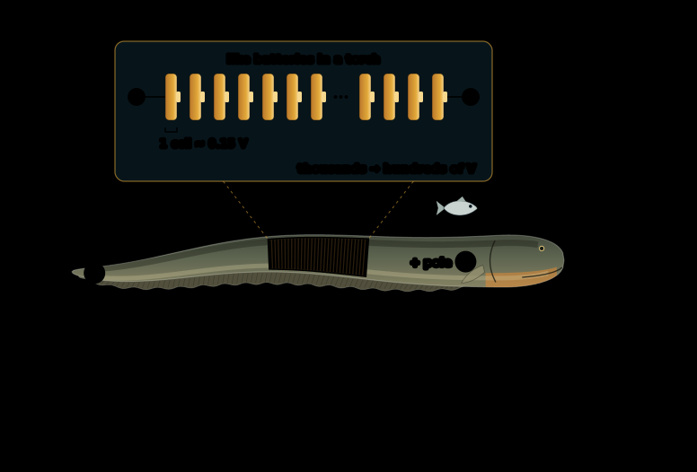
How does a fish produce hundreds of volts? The answer lies in a principle every torch uses: many small batteries in a row. The lenses below follow it – from ions crossing a cell membrane to the circuit that forms when an eel leaps at a threat.
The attack in four steps
Electric eels give off two kinds of electric pulses, each a head-positive pulse lasting one to two milliseconds: a weak one for sensing and a strong one, up to hundreds of volts, emitted in volleys of up to 500 pulses per second 4 5. With high-speed video and recordings of eel and prey at the same time, Catania found that eels use the strong pulses in a remarkably refined way 6 4:
- Locate. A resting eel gives off weak pulses at a low rate; an excited, hunting eel can emit them at 10–20 Hz, as Catania reports from earlier work. These pulses serve for active electroreception 4: the eel senses objects by how they distort its own electric field. Prey that hides is still a problem.
- Flush out. When prey is hidden, the eel can emit periodic volleys of two or three strong discharges. They cause a massive involuntary twitch in the prey and so reveal its location 6. The eel detects the resulting water movement with its mechanoreceptors and answers 20–40 ms later with a full volley and a strike 4.
- Immobilise. In a full high-voltage volley, each pulse typically activates the motor neurons of nearby prey – the nerve cells that make muscles contract. The prey freezes in a whole-body contraction, a tetanus, and cannot escape: the eel remote-controls its muscles 6 4. In high-speed videos, all voluntary movement of a fish stopped within 3–4 ms of the first pulse 4.
- Exhaust. Large or struggling prey gets a special treatment. The eel grips it and often curls to bring its tail to the opposite side, which more than doubles the strength of the electric field in the prey. Then it delivers repeated volleys of pulses at about 100 Hz, which fatigue the prey’s muscles – a strategy Catania compares to a venom that paralyses muscles 5.
Biology lens: one genus, three species, six inventions
Three species with different homes. De Santana and colleagues examined 107 electric eels from across Greater Amazonia. Shields are old, geologically stable cores of the continent. Electrophorus electricus is restricted to the Guiana Shield in the north; E. voltai lives in the rivers of the Brazilian Shield to the south and in some rivers of the Guiana Shield; E. varii lives in the lowland floodplains and streams of the Amazon Basin. Shield rivers never run low in oxygen, have rocky beds and rapids and low electrical conductivity – below 30 µS/cm – whereas lowland waters range from low-conductivity blackwaters to high-conductivity whitewaters and include floodplains that are seasonally low in oxygen. The three lineages split during the Miocene and Pliocene; E. voltai and E. electricus, for example, an estimated 3.6 million years ago (95 % interval 4.7–2.5 million years). Outwardly, the species are hard to tell apart; they differ in the shape of skull and head and in their genes 3. Electric eels can grow up to 2.5 m long, as the authors note 3.
Three organs, one body. An electric eel has three electric organs, called the main organ, Hunter’s organ and Sachs’ organ 2. In all three species, the weak pulses come from Sachs’ organ and the rear part of Hunter’s organ, the strong ones from the main organ and the front part of Hunter’s organ 3. The cells of these organs, the electrocytes, develop from muscle precursor cells but are notably larger than muscle fibres and lack the machinery for contraction 2. They span the rear 80 % of the eel’s body 1.
Six independent inventions. Electric organs evolved at least six times independently in fishes. Jason Gallant and colleagues sequenced the genome of the electric eel and compared electric organs and muscles in three lineages that evolved them separately. Despite millions of years of evolution and very different cell shapes, these lineages used similar transcription factors – proteins that switch genes on and off – and similar developmental and cellular pathways to build their electric organs 2. This is convergent evolution: the same solution reached on different paths.
A leap that confirms Humboldt. In March 1800, Humboldt watched fishermen in South America “fish with horses”: they drove horses into a pool with electric eels, which attacked by pressing themselves against the horses while discharging. Once the eels were exhausted, they could be collected safely. Later investigators were sceptical of the story. In 2016, Catania reported a defensive behaviour that supports it: when a large, partially submerged conductor approached, eels leaped out of the water, pressed their chin against it and discharged high-voltage volleys 7. The behaviour appeared to be ubiquitous in eels over 60 cm in his aquariums 7. Catania thinks the leap helps eels defend themselves in the dry season, when they can be trapped in small pools and cannot retreat; eels are obligate air breathers, well suited to such pools that often run low in oxygen, as he summarises 7.
Physics lens: batteries in series, current through the water
Voltages add in series. The electrocytes of the eel’s electric organ are flat and asymmetric, and they lie in a row. Their voltages add up, “much like batteries stacked in series in a flashlight” 2. Each firing electrocyte contributes about 0.15 V. Large electric eels stack thousands of electrocytes in series and reach more than 600 V; arranging many stacks in parallel – like several rows of batteries side by side – makes peak currents approaching 1 A possible at short circuit 1. Series raises the voltage, parallel lowers the internal resistance and raises the current.
A dipole in the water. During a discharge, the head is the positive pole and the tail the negative one. Current flows from the head through the water back to the tail and forms a roughly dipole-shaped field around the eel 4 – the same shape as the field of a bar magnet. A fish near the head feels mostly the positive pole. When the eel curls and brings its tail to the other side of the prey, the two poles act together: the prey is sandwiched between them, and the field inside it can more than double 5. The eel cannot raise the amplitude of its pulses during a volley – every electrocyte already fires – so curling simply focuses the fixed output on the prey, “much like focusing the fixed power of a flashlight into a smaller area” 4.
Why water is a problem for defence. Water conducts, so most of the current spreads through the water around the eel. Faraday already noticed in 1838 that when he touched an eel with one hand in the water, the shock was felt only in that hand and was not very strong 7. A land animal with only its legs in the water may get only a small part of the current 7.
The leap as a short circuit. In the leap, the eel presses its chin against the threat. This short-circuits its electric organ through the threat, and as the eel rises higher, more and more power is diverted to it 7. The proposed reason is the path back to the water: the higher the eel rises, the longer – and presumably the more resistant – the path from its head along its body down to the water, so a larger share of the current takes the path through the threat 7 4. In measurements, voltage and current in the target increased dramatically as the eels ascended 7.
What a human feels. Catania measured the current that flowed through a human arm – apparently his own, as he later hinted 5 – during the leap of a comparatively small eel: it peaked at 40–50 mA, far above the thresholds for activating nociceptors – the receptors that signal pain – in humans and horses. No involuntary tetanus was felt, and the unpleasant sensations stayed in the affected arm. The main purpose of the leap, Catania suggests, is to deter predators by briefly causing intense pain 8.
Mathematics lens: the eel as a battery with internal resistance
A battery model. Physiologists analyse electrocytes with the same methods as batteries 4. A real battery has an electromotive force ε – the voltage it produces with no current flowing – and an internal resistance r. When a current I flows, part of the voltage is lost inside the battery, and the voltage V at its terminals drops:
This is how Catania measured ε and r of living eels: an adjustable resistor is put into the circuit, V and I are measured for each high-voltage pulse, and the points form a straight line whose slope is −r 4. For five eels of different sizes, Catania found – as read from Fig. 12 of his 2019 review, which shows the data of his 2017 studies – electromotive forces between 179 and 382 V and internal resistances between 438 and 960 Ω; as an eel grows and adds electrocytes, Catania writes, its electromotive force increases and its internal resistance decreases – a trend rather than a strict rule: in his figure, the largest eel has a slightly higher internal resistance (450 Ω) than the next smaller one (438 Ω) 4.
The circuit of the leap. For a small eel – the top one in Catania’s figure of the five – the values in the same Fig. 12 were ε = 198 V and r = 960 Ω; the water path back to the tail had about 400 Ω, and the return path from the head down to the water reached about 2,400 Ω at the top of the leap. A human arm, the target, had about 2,100 Ω 4. The return path Ro and the target R are connected in parallel: both lead from the eel’s head back to the water. Catania compares the return path to a volume control: the higher the eel leaps, the larger Ro, and the “louder” the attack 4.
One battery seen from the target. Any such network of a source and resistors looks, from the target’s point of view, like a single battery with a voltage Vth and an internal resistance Rth – its Thévenin equivalent. Here (a short calculation for this article):
With the values above, the target sees 126 V behind 868 Ω, and the arm gets I = 126 V / (868 Ω + 2,100 Ω) ≈ 43 mA – the value Catania’s Fig. 12 gives for this circuit 4. This agreement is a consistency check, not an independent result: Catania calculated the arm resistance from the measured current. As Ro grows, Vth rises towards the full ε: this is why a higher leap means a stronger shock.
When does the target get the most power? The power in the target is
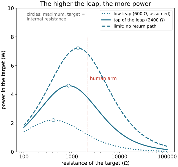
A target with a very small resistance gets a large current but little voltage; one with a very large resistance gets voltage but almost no current. In between lies a maximum. Setting the derivative dP/dR to zero gives R = Rth and Pmax = Vth2 / (4 Rth) – the maximum power transfer theorem: a source delivers the most power into a load whose resistance equals its own internal resistance. For the small eel, that would be a target of 868 Ω receiving 4.6 W; the arm, with 2,100 Ω, receives 3.8 W – peak power during each pulse, because Catania’s values are peak values (a calculation for this article; the computer science lens shows the whole curve).
Chemistry lens: ion gradients as fuel
Chemical energy stored as a gradient. Every animal cell keeps more potassium ions (K+) inside and more sodium ions (Na+) outside. This uneven distribution – an ion gradient – stores energy, like water held behind a dam. The enzyme Na+/K+-ATPase, the sodium–potassium pump, builds the gradient by using the energy of ATP to move sodium out and potassium in; in the electric organ it rebuilds the gradients after a discharge 1. In the eel’s electrocytes, the sodium pumps are concentrated on the invaginated, non-innervated face of the cell 2.
Two faces, one voltage. An electrocyte is a flat cell with two different faces. At rest, open potassium channels on both faces give each membrane a potential of −85 mV; they point in opposite directions and cancel out, so the cell as a whole produces no voltage. When a nerve signal arrives at the innervated, rear face, voltage-gated sodium channels there open and potassium channels close. This face now produces an action potential of +65 mV, while the front face stays at −85 mV. Together, the cell now has about 150 mV across it 1. Because the front face is permeable to potassium and the rear face to sodium, the electrocyte taps two opposing gradients of two different ions at once with low-resistance salt solutions on both sides – a non-obvious strategy to maximise power output, as Thomas Schroeder and colleagues point out 1.
Acetylcholine: from nerve to cell. The nerve endings on the innervated face are cholinergic 2: they release the signalling molecule acetylcholine, which opens receptors on the electrocyte. The electric eel was used to isolate this acetylcholine receptor in the 20th century 4. The same kind of receptor plays a role in the hunt: curare, an arrow poison, blocks the acetylcholine receptors of muscles. When Catania injected curare into a pithed fish, the eel’s pulses no longer made its muscles contract – proof that the eel acts on the motor neurons, not directly on the muscles 5.
Weak and strong with the same cells. For a weak pulse, an action potential reaches every electrocyte, but only a subset with a low threshold fires. For a strong pulse, the nerves fire at a very high rate, and the signals add up until every electrocyte fires, as Catania summarises 4. All electrocytes along the roughly 1 m long organ must fire within the about 2 ms of a discharge. Since nerve signals do not travel fast enough for this, the eel slows down the arrival of signals at the parts of the organ closest to the nerve centre that commands the discharge 1.
Better than nature? Jian Xu and David Lavan calculated how the ion channels and pumps of an eel electrocyte turn ion gradients into action potentials. Using the parameters of this model, they designed – as a calculation, not a built device – an artificial cell from an optimised selection of these conductors. In their calculation, it has a higher power density and a greater energy conversion efficiency than the electrocyte; they suggest that such artificial cells could power medical implants and other small devices 9.
Computer science lens: simulating the leap
How much current and power does a leaping eel push through a threat, and how does that depend on the target and on the height of the leap? The program below models the eel as a battery with internal resistance and computes current and power in the target with the Thévenin equivalent from the mathematics lens. Inputs and assumptions: the voltage of one electrocyte, 0.15 V 1; the measured electromotive force of 198 V and internal resistance of 960 Ω of a small eel, one of five, the water path of about 400 Ω, the return path of about 2,400 Ω at the top of the leap and the arm resistance of about 2,100 Ω 4. A return path of 600 Ω stands for a lower point of the leap; it is an assumption, as is the limit of a return path with practically infinite resistance.
import numpy as np
import matplotlib.pyplot as plt

# How much power does a leaping electric eel push through a threat? An equivalent circuit.
CELL_EMF = 0.150         # voltage of one firing electrocyte (V), Schroeder et al. 2017
# Circuit of a small eel, measured or estimated by Catania (summarised in Catania 2019, Fig. 12):
EMF, R_INTERNAL = 198.0, 960.0   # electromotive force (V) and internal resistance (Ω)
R_WATER = 400.0          # resistance of the water path back to the tail (Ω)
RETURN_PATH = 2400.0     # head-to-water path along the eel at the top of the leap (Ω)
R_ARM = 2100.0           # a human arm, the target (Ω)
ROWS = 1                 # how many copies of the eel's organ work in parallel (1 = the real eel)


def thevenin(emf, r_source, r_return):
    """Seen from the target, the eel, the water and the return path act like one battery:
    the return path divides the voltage and sits in parallel with the target."""
    r_loop = r_source + R_WATER
    v_th = emf * r_return / (r_loop + r_return)
    r_th = r_loop * r_return / (r_loop + r_return)
    return v_th, r_th


def target_power(r_target, v_th, r_th):
    """Current and power in a target of resistance R: I = V/(R_th + R), P = I²·R."""
    current = v_th / (r_th + r_target)
    return current, current**2 * r_target


r_eel = R_INTERNAL / ROWS                 # rows in parallel divide the internal resistance
for volts in (EMF, 600.0, 860.0):
    print(f"{volts:.0f} V needs about {volts / CELL_EMF:,.0f} electrocytes in series")

v_th, r_th = thevenin(EMF, r_eel, RETURN_PATH)
print(f"seen from the target: {v_th:.0f} V behind {r_th:.0f} Ω")
i_arm, p_arm = target_power(R_ARM, v_th, r_th)
i_eel = EMF / (r_eel + R_WATER + RETURN_PATH * R_ARM / (RETURN_PATH + R_ARM))
print(f"current leaving the eel: {1e3 * i_eel:.0f} mA")
print(f"arm ({R_ARM:.0f} Ω): {1e3 * i_arm:.0f} mA, {p_arm:.1f} W")
print(f"best target: {r_th:.0f} Ω gets {target_power(r_th, v_th, r_th)[1]:.1f} W (maximum power transfer)")
for r_return, name in ((600.0, "low leap"), (RETURN_PATH, "top of the leap"), (1e9, "no return path")):
    i, p = target_power(R_ARM, *thevenin(EMF, r_eel, r_return))
    print(f"{name}: arm current {1e3 * i:.0f} mA, {p:.1f} W")

plt.rcParams["font.size"] = 13      # large type: the chart stays readable on a phone
fig, ax = plt.subplots(figsize=(6.4, 6.0), layout="constrained")
r_target = np.logspace(2, 5, 400)
for r_return, label, style in ((600.0, "low leap (600 Ω, assumed)", ":"),
                               (RETURN_PATH, "top of the leap (2400 Ω)", "-"),
                               (1e9, "limit: no return path", "--")):
    v, r = thevenin(EMF, r_eel, r_return)
    ax.plot(r_target, target_power(r_target, v, r)[1], ls=style, lw=2.5, color="#1f6f8b", label=label)
    ax.plot(r, target_power(r, v, r)[1], "o", ms=8, mfc="white", color="#1f6f8b")
ax.axvline(R_ARM, ymax=0.8, color="#c0392b", lw=1.5, ls="-.")
ax.text(R_ARM * 1.15, 5.0, "human arm", color="#c0392b", fontsize=12)
ax.text(115, 9.7, "circles: maximum, target =\ninternal resistance", fontsize=11, color="dimgrey", va="top")
ax.set_xscale("log")
ax.xaxis.set_major_formatter("{x:g}")
ax.set_xlabel("resistance of the target (Ω)")
ax.set_ylabel("power in the target (W)")
ax.set_ylim(0, 10)
ax.set_title("The higher the leap, the more power")
ax.legend(loc="upper right", fontsize=11)
plt.show()
Output · what the code prints, re-run at every build
198 V needs about 1,320 electrocytes in series
600 V needs about 4,000 electrocytes in series
860 V needs about 5,733 electrocytes in series
seen from the target: 126 V behind 868 Ω
current leaving the eel: 80 mA
arm (2100 Ω): 43 mA, 3.8 W
best target: 868 Ω gets 4.6 W (maximum power transfer)
low leap: arm current 24 mA, 1.2 W
top of the leap: arm current 43 mA, 3.8 W
no return path: arm current 57 mA, 6.9 W
What the result teaches:
- The model matches the measurement. With Catania’s values, the program gives 43 mA through the arm and 80 mA leaving the eel – the same as the currents of about 0.043 A and 0.08 A that Fig. 12 of Catania’s review shows for this circuit 4. This is a consistency check of the circuit, not an independent measurement: the arm resistance was itself calculated from the measured current.
- Higher leap, stronger shock. At a lower point of the leap (return path 600 Ω, assumed), the arm gets 24 mA and 1.2 W; at the top of the leap 43 mA and 3.8 W; in the limit of no return path, 57 mA and 6.9 W. This is the “volume control” of the leap.
- There is a best target. At the top of the leap, a target of 868 Ω would get the most power, 4.6 W. The arm, with 2,100 Ω, receives 3.8 W – more than four fifths of this maximum.
- Thousands of cells in a row. At 0.15 V per cell, 198 V needs about 1,320 electrocytes in series, 600 V about 4,000 and 860 V about 5,733 – a rough estimate, because it assumes that all cells fire with exactly 0.15 V and that no voltage is lost inside the eel.
Try it yourself:
- Give the eel twice as many rows of cells in parallel: set
ROWS = 2. The internal resistance halves, and the arm current rises from 43 to 53 mA and the power from 3.8 to 5.9 W; the best target now has 644 Ω. - Put in the eel with the highest voltage of Catania’s five, 382 V and 450 Ω: change
198.0, 960.0to382.0, 450.0. Assuming the same water and return path, the arm current rises to 103 mA and the power to 22.5 W – an assumption, because for this eel no leap was measured with an arm.
Tested output with ROWS = 2
198 V needs about 1,320 electrocytes in series
600 V needs about 4,000 electrocytes in series
860 V needs about 5,733 electrocytes in series
seen from the target: 145 V behind 644 Ω
current leaving the eel: 99 mA
arm (2100 Ω): 53 mA, 5.9 W
best target: 644 Ω gets 8.1 W (maximum power transfer)
low leap: arm current 33 mA, 2.2 W
top of the leap: arm current 53 mA, 5.9 W
no return path: arm current 66 mA, 9.3 W
Tested output with 382.0, 450.0
382 V needs about 2,547 electrocytes in series
600 V needs about 4,000 electrocytes in series
860 V needs about 5,733 electrocytes in series
seen from the target: 282 V behind 628 Ω
current leaving the eel: 194 mA
arm (2100 Ω): 103 mA, 22.5 W
best target: 628 Ω gets 31.7 W (maximum power transfer)
low leap: arm current 64 mA, 8.7 W
top of the leap: arm current 103 mA, 22.5 W
no return path: arm current 129 mA, 35.2 W
Physical AI lens: soft batteries and robots with an electric sense
Physical AI – embodied artificial intelligence: machines that sense and act in the physical world – needs two things the eel has: a power source that works inside a soft, wet body, and senses that work where eyes fail. Engineers are learning from the eel and its weakly electric relatives for both.
An artificial electric organ. Power sources inside living organisms should be biocompatible, mechanically flexible and able to use the chemical energy available in the body – criteria conventional batteries were not designed for. Schroeder and colleagues built an eel-inspired power source from hydrogels – soft, water-filled polymer networks. Like an electrocyte, each repeating unit consists of four gels: a salty and a less salty gel separated by gels that let only positive or only negative ions pass. Each of these “gel cells” produced 130–185 mV, comparable to a single electrocyte, and 2,449 gels in series added up to 110 V. To make all compartments touch at the same moment, as the eel fires all electrocytes together, the gels were printed on two sheets and pressed together, or stacked with a folding pattern developed for solar panels in space. Unlike typical batteries, the systems are soft, flexible, transparent and potentially biocompatible 1.
Still far from the eel. The power was modest: 50 µW across 2,449 gels in the printed version, and even the best folded geometry reached a power density 2–3 orders of magnitude lower than the eel’s electrocyte layers. Thinner and more selective membranes could close part of the gap. The authors suggest that artificial electric organs might one day power implants such as pacemakers, implantable sensors or prostheses, and serve as deformable power sources in soft robots 1.
Seeing with electric fields. Many electric fishes sense their surroundings with weak electric fields, and the eel even uses its strong pulses to track fast-moving prey 4. James Solberg, Kevin Lynch and Malcolm MacIver built a robot that finds objects underwater the same way: it moves an emitter and sensors that create and measure an electric field, inspired by active electrolocation in two groups of weakly electric freshwater fishes. A particle filter – a probabilistic algorithm – keeps an estimate of where the target is and refines it with each measurement. The robot located targets in low-conductivity water, where electric fishes live, and also in water of ocean salinity, where electrolocation is not observed in nature 10.
What we still do not know
Electric eels have been studied for centuries, yet basic questions are open. Little is known about what they eat in the wild, whether other strongly electric fishes also activate the motor neurons of their prey, and how an eel protects its own nervous system from its discharges 4. The three species have only just been described: field studies of their discharges, the genomes of all three species and samples from regions such as the upper Rio Negro and the Orinoco may reveal more – including further diversity within the genus 3.
Artificial electric organs are just approaching the power needed by the lowest-power implants. If future designs narrow the gap to the natural organ by one or two orders of magnitude, they may one day power implants, wearables and other mobile devices with metabolically available energy 1 – the eel’s principle of many small voltages in a row, rebuilt in a soft material.
Frequently asked questions
Is the electric eel really an eel?
No. Despite its name, the electric eel is not a true eel but a South American knifefish (order Gymnotiformes), related to other electric fishes of South America. Since 2019, three species are recognised: Electrophorus electricus, Electrophorus voltai and Electrophorus varii.
How many volts can an electric eel produce?
The highest value reported, in 2019, is 860 volts, from an Electrophorus voltai about 1.2 metres long, measured out of the water from snout to tail. Before, about 650 volts was the figure cited for electric eels. In Catania’s data, the voltage tends to grow with the size of the eel: in five eels of different sizes, Kenneth Catania measured electromotive forces between 179 and 382 volts.
How does an electric eel make electricity?
With cells called electrocytes, which evolved from muscle cells. At rest, the two faces of a flat electrocyte carry equal and opposite voltages, so the cell produces nothing. When a nerve signal arrives, sodium channels open on one face only, and the cell suddenly has about 0.15 volts across it. Thousands of electrocytes lie in series, like batteries in a torch, so their voltages add up.
Why do electric eels jump out of the water?
To defend themselves. In the water, an eel’s current spreads out, and an animal with only its legs in the water may get only a small part of it. When the eel leaps up a threat with its chin pressed against it, more and more of the current flows through the threat the higher it rises. The shock is very painful; the leap probably serves to deter predators, for example when eels are trapped in pools in the dry season.
Why doesn’t an electric eel shock itself?
That is still an open question. Kenneth Catania lists how an eel protects its own nervous system from its high-voltage discharge among the questions that remain to be investigated.
Sources
- Schroeder, T. B. H., Guha, A., Lamoureux, A., VanRenterghem, G., Sept, D., Shtein, M., Yang, J., Mayer, M. (2017). An electric-eel-inspired soft power source from stacked hydrogels. Nature 552, 214–218. doi:10.1038/nature24670 · Open Access
- Gallant, J. R., Traeger, L. L., Volkening, J. D., Moffett, H., Chen, P. H., Novina, C. D., Phillips, G. N., Anand, R., Wells, G. B., Pinch, M., Güth, R., Unguez, G. A., Albert, J. S., Zakon, H. H., Samanta, M. P., Sussman, M. R. (2014). Genomic basis for the convergent evolution of electric organs. Science 344, 1522–1525. doi:10.1126/science.1254432 · Open Access
- de Santana, C. D., Crampton, W. G. R., Dillman, C. B., Frederico, R. G., Sabaj, M. H., Covain, R., Ready, J., Zuanon, J., de Oliveira, R. R., Mendes-Júnior, R. N., Bastos, D. A., Teixeira, T. F., Mol, J., Ohara, W., Castro e Castro, N. C., Peixoto, L. A., Nagamachi, C., Sousa, L., Montag, L. F. A., Ribeiro, F., Waddell, J. C., Piorsky, N. M., Vari, R. P., Wosiacki, W. B. (2019). Unexpected species diversity in electric eels with a description of the strongest living bioelectricity generator. Nature Communications 10, 4000. doi:10.1038/s41467-019-11690-z · Open Access
- Catania, K. C. (2019). The Astonishing Behavior of Electric Eels. Frontiers in Integrative Neuroscience 13, 23. doi:10.3389/fnint.2019.00023 · Open Access
- Catania, K. C. (2021). Electric Eels Wield a Functional Venom Analogue. Toxins 13, 48. doi:10.3390/toxins13010048 · Open Access
- Catania, K. (2014). The shocking predatory strike of the electric eel. Science 346, 1231–1234. doi:10.1126/science.1260807
- Catania, K. C. (2016). Leaping eels electrify threats, supporting Humboldt’s account of a battle with horses. Proceedings of the National Academy of Sciences 113, 6979–6984. doi:10.1073/pnas.1604009113 · Open Access
- Catania, K. C. (2017). Power Transfer to a Human during an Electric Eel’s Shocking Leap. Current Biology 27, 2887–2891.e2. doi:10.1016/j.cub.2017.08.034
- Xu, J., Lavan, D. A. (2008). Designing artificial cells to harness the biological ion concentration gradient. Nature Nanotechnology 3, 666–670. doi:10.1038/nnano.2008.274 · Open Access
- Solberg, J. R., Lynch, K. M., MacIver, M. A. (2007). Robotic Electrolocation: Active Underwater Target Localization with Electric Fields. Proceedings 2007 IEEE International Conference on Robotics and Automation , 4879–4886. doi:10.1109/robot.2007.364231
Text and figures: CC BY-SA 4.0. Found a mistake? Open an issue or edit this article on GitHub.